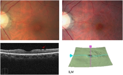
Figure 1.
Upper left: preoperative fundus photography. No retinal nerve fiber layer defect in the perimacular area. Upper right: 12-months follow-up images after vitrectomy. Multiple dark round lesions in the superior temporal perimacular area. Lower left: well-demarcated DONFL (arrow) margin on OCT (vertical axis). Lower right: three-dimensional OCT mapping shows multiple round DONFLs, with a ‘beaten bronze' cobblestone-like appearance, in the superior temporal perimacular area.

