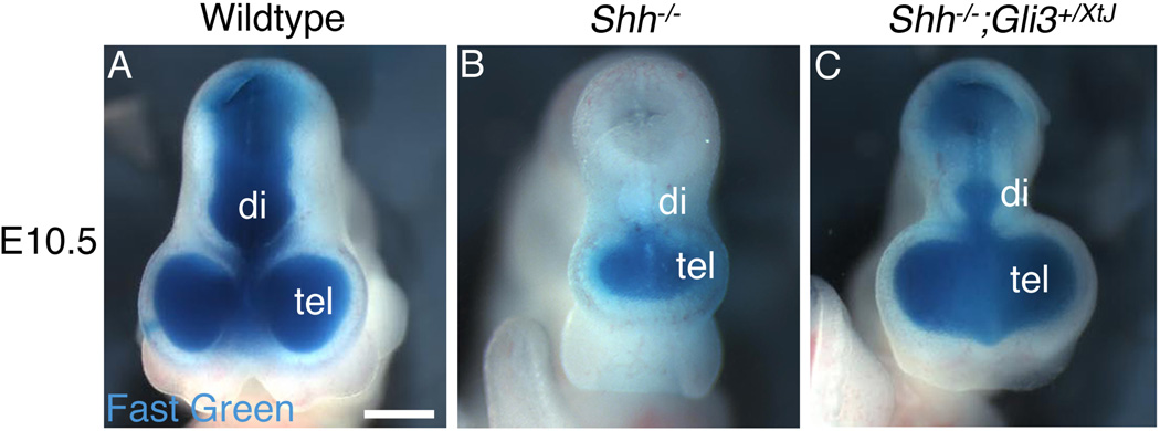
Figure 1. Blockage of the third ventricle is a feature of HPE in Shh null embryos and is rescued by removing one functional copy of Gli3.
The telencephalic vesicle (tel) in E10.5 embryos was injected with a solution of fast green to fill the ventricular system, with a hole cut in the mesencephalon to allow the dye to exit. The lateral ventricles and third ventricle are filled with dye in a wildtype embryo (A). Ventricular flow is blocked at the level of the third ventricle in a Shh null embryo, so that the forebrain ventricular system forms a sealed monoventricle (B). In contrast, ventricular system continuity is restored in a Shh−/−;Gli3+/XtJ embryo (C), although the diencephalic ventricle (di) is reduced in size compared with wildtype. Scale bar is 700µm.

