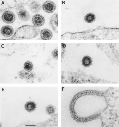
FIG. 2.
Analysis of structure of cell-associated virions by electron microscopy. Cells producing wild-type or mutant particles were fixed and sectioned for electron microscopy. (A) Wild type; (B) PR−; (C) S1KK; (D) S2D; (E) S3R; (F) S3KK. The samples shown in panels A to E were processed by the tannic acid procedure, while that in panel F is a conventional thin-section electron micrograph. Scale bar, 100 nm.

