
Figure 3.
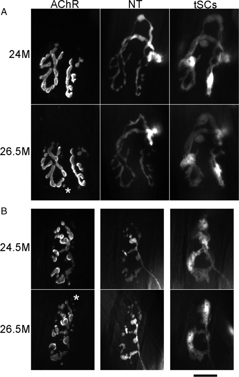
Most NMJs in aging muscles, regardless of whether they have the young or the old phenotype, are stable over time. Images were obtained over the indicated time intervals from the same NMJs. Labeling is as described in Figure 1. An image of AChRs was made during the second imaging of each NMJ (bottom row in each panel) before any additional Alexa 647 bungarotoxin was applied. This image therefore shows the receptors persisting from the previous imaging session. Images of AChRs here and in Figure 4 were captured and are presented at the same camera gain and exposure time so that the intensity differences represent those seen in the microscope. A, Young-appearing junction that showed no change and persistent, strong labeling of AChRs from the first imaging session. An additional four images collected between 24 and 26.5 months showed no change. B, Fragmented, old-appearing junction that showed no change and persistent strong labeling of AChRs from the previous imaging session. An additional three images between 24.5 and 26.5 showed no change. The asterisks in the two images mark one of several examples where AChRs, labeled in the prior imaging session, appear as spots of label that are now internal to the fiber (Akaaboune et al., 1999). Scale bar, 20 μm.