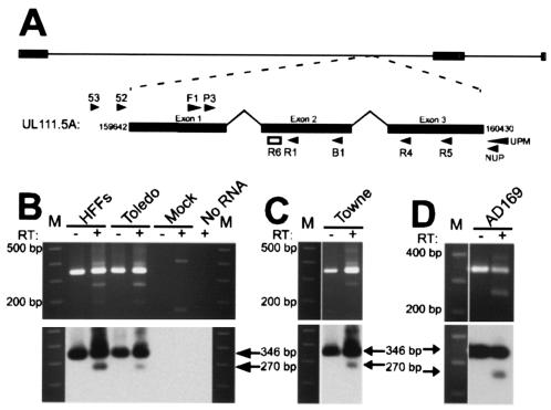
FIG. 1.
(A) Schematic representation of the CMV genome with the UL111.5A transcript expanded to show the position of primers (arrow heads) and probe (open box) used in the present study. (B) RT-PCR analysis of UL111.5A region gene expression in latently infected GM-Ps. Detection of UL111.5A region transcripts in CMV stain Toledo productively infected HFFs (day 4 postinfection) and latently infected GM-Ps (day 14 postinfection) (B) and CMV strain Towne (C) and AD169 (D) latently infected GM-Ps (day 14 postinfection). Panels B to D show ethidium bromide-stained agarose gels (upper panel) and the corresponding Southern blots (lower panel) of products generated from RT-PCR analysis for UL111.5A region transcripts. The sizes of the RT-PCR products are indicated with arrows and the numbers to the left of the gels indicate the size of the adjacent molecular weight size markers (M). The presence (+) or absence (−) of reverse transcriptase (RT) in the reaction mixture is indicated.

