
Figure 5.
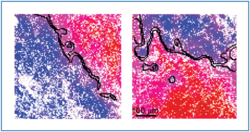
Automated tumor margin identification (black curves) overlaid on 2 smoothed representative SR-NIVI images. The 2 boundaries of the margin demarcate normal and tumor domains at the greater than 99% confidence interval. Tumor margins are readily resolved to 100 μm.