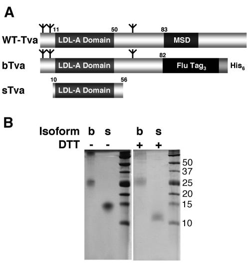
FIG. 1.
Tva isoforms. (A) Schematic diagram of Tva-derived proteins used in this study. Numbers refer to the positions of residues in mature wild-type (WT) Tva. MSD, membrane-spanning domain. Flu Tag3, three copies of an influenza virus HA epitope tag. Branched symbols denote positions of N-linked glycosylation sites. (B) Coomassie R-250-stained gels of purified bTva and sTva. Where noted, samples were boiled in 100 mM DTT prior to loading.

