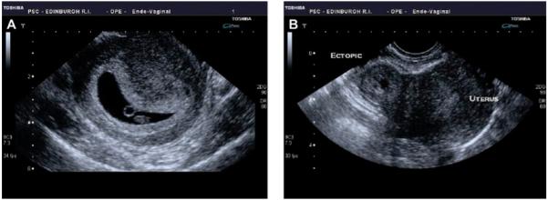
Figure 1.

Transvaginal ultrasound images of an intrauterine pregnancy (IUP) and ectopic pregnancy. (A) An IUP at 6 weeks. The central dark area is the intrauterine gestational sac and within the sac is a circular ringed structure that is the yolk sac. The small oval structure below the yolk sac is the fetus. (B) An ectopic pregnancy. To the right of the image is the normal uterus and to the left of the uterus is the doughnut-shaped ectopic pregnancy.
