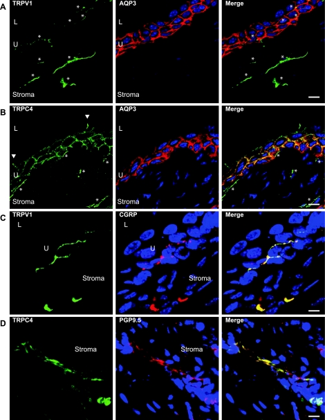
Fig. 3.
Immunofluorescent localization of TRP channels in and near urothelium (U). Multicolor confocal laser scanning immunofluorescence shows the cellular locations of transient receptor potential (TRP) family members TRPV1 (A and C) and TRPC4 (B and D). Antibodies to aquaporin-3 (AQP3) were used to define the intermediate and basal urothelial cell borders (red in A and B), while TRP immunostaining is green. TRP-positive fibers are indicated by asterisks, and the TRPC4-positive lateral boundaries of an umbrella cell are highlighted by arrowheads. Right panels (C and D) show colocalization of both TRPV1 and TRPC4 (green) with neuronal marker proteins calcitonin gene-related peptide (CGRP) and protein gene product 9.5 (PGP9.5; red), indicating that TRP-positive fibers are neurons. Scale bar = 10 μm. L, lumen; U, urothelium. Figure is modified from Ref. 79.

