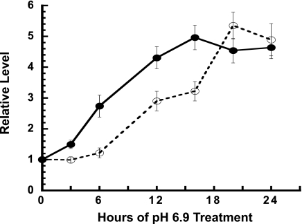
Fig. 1.
pH-responsive increase in PEPCK protein and mRNA in LLC-PK1-F+-9C cells. Cells were grown to confluence in normal medium and then either maintained in normal (pH 7.4) medium or transferred to acidic (pH 6.9) medium for the indicated times. All of the cells were harvested at the same time and quantified for PEPCK and β-tubulin protein by Western blotting and for PEPCK and GAPDH mRNAs by real-time RT-PCR. The ratio of PEPCK protein to β-tubulin was normalized to the average of the control samples and used to calculate the fold increase in PEPCK (o—o). The PEPCK mRNA data were similarly standardized vs. the levels of GAPDH mRNA (•-•). The data are plotted as means ± SE of triplicate samples.

