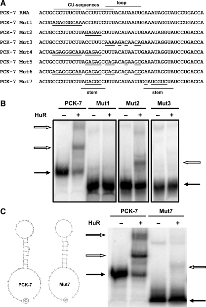
Fig. 8.
Recombinant HuR binds to 2 distinct sites within the PCK-7 RNA. A: schematic diagram lists the sequences of the PCK-7 RNA and the Mut1 through Mut7 constructs. It also illustrates the regions that contain the CU sequences and the stem and loop sequences. The nucleotides modified in the 7 different mutated constructs of PCK-7 RNA are underlined. B: electrophoretic mobility shift assays were performed using a constant concentration of recombinant HuR and equivalent amounts of [32P]-labeled PCK-7 RNA or the Mut1, Mut2, or Mut3 RNAs. The panel was constructed from 3 separate gels, each of which used the labeled PCK-7 RNA as a positive control. C: schematic diagram illustrates the secondary structure of the PCK-7 RNA and PCK-7 Mut7 RNA as predicted by RNAdraw. An electrophoretic mobility shift assay was performed using a constant concentration of recombinant HuR and equivalent amounts of [32P]-labeled PCK-7 RNA or the PCK-7 Mut7 RNA. Samples were run on 5% native polyacrylamide gels. The solid arrows indicate the unbound RNA and the outlined arrows indicate the RNA:protein complexes.

