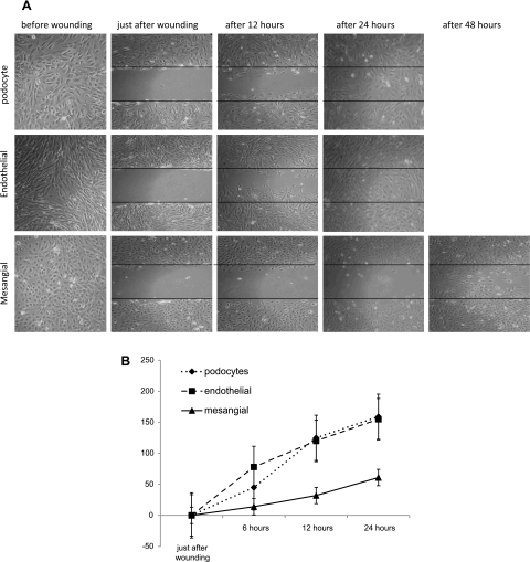
Fig. 6.
A: wound healing assay. Podocytes, glomerular endothelial, and mesangial cells were seeded at confluence into 6-well culture plates and allowed to adhere overnight. Then, the medium was altered to 0.2% FCS to reduce cell proliferation. Two days afterward, wounds were made. Images from 2 marked fields/well were taken under phase-contrast examination, just before wounding, immediately after wounding (0), and after 12, 24, and 48 h postwounding. Wounds in podocytes and endothelial cells wells were completely closed after 24 h, and unexpectedly in mesangial cells the wound was not completely closed even after 48 h. B: proportion of cells invading the scratch-made wound at different time points (after 6, 12, and 24 h); unexpectedly, mesangial cells migrate more slowly than podocytes and endothelial cells.

