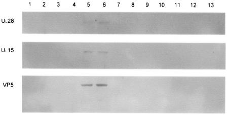
FIG. 3.
Immunoblots of B capsids purified through two sequential 20- to-50% sucrose gradients. The 14-ml gradient was fractionated from the top (fraction one), and the proteins present in each fraction were separated on an 8% denaturing polyacrylamide gel before being transferred to a PVDF membrane. The membrane was then probed with antisera against UL28, UL15, or VP5 and developed using the ECL+ method (see Materials and Methods). The image was generated using a Molecular Dynamics Storm PhosphorImager with chemiluminescence detection capability.

