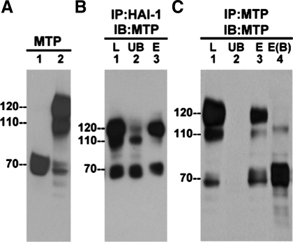
Fig. 1.
Formation of non-heptatocyte activator inhibitor (HAI)-1 matriptase (MTP) complexes in mammary epithelial cells. A: 184 A1N4 mammary epithelial cells were exposed to basal medium (lane 1) or a pH 6.0 buffer (lane 2) for 20 min to induce MTP activation. Cell lysates were prepared and analyzed for MTP by blotting with the MTP monoclonal antibody (mAb) M24. B: cell lysates were immunoprecipitated (IP) using HAI-1 mAb M19 beads (IP:HAI-1), and the captured proteins were eluted using pH 2.4 glycine buffer. The lysate (L), the unbound fraction (UB), and the eluted fraction (E) were analyzed by immunoblot for MTP with mAb M24 (IB:MTP). C: cell lysates were immunoprecipitated by using MTP mAb 21–9 beads and the bound proteins eluted with pH 2.4 glycine buffer (IP:MTP). Proteins samples from the lysate (L), the unbound fraction (UB), and the eluted fractions (E) were incubated with SDS sample buffer at room temperature (lane 1 through lane 3) or at 95°C (lane 4, B) for 5 min before electrophoresis and analyzed by immunoblotting for MTP with the mAb M24 (IB:MTP).

