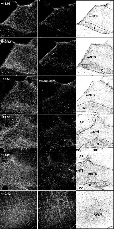
Fig. 1.
Serotonin immunoreactivity in five coronal planes of the nucleus of the solitary tract (nTS) (5 sets of images on top) and one coronal plane of the rostroventrolateral medulla (RVLM) (set of images on bottom) from brains of representative rats treated with vehicle (left) or 5,7-dihydroxytryptamine (middle) in the caudal dorsomedial brain stem. Approximate location of the section (in mm caudal to bregma) is indicated in the top left corner of the column of images on the left. Images in each row are from the same approximate location. Column of images on the right is the inverse of the column of images on the left showing the location of borders drawn for quantification of specific areas of interest: medial nTS (mNTS), dorsal motor nucleus of the vagus (X), area postrema (AP), commissural nTS (cNTS), and RVLM. CC, central canal.

