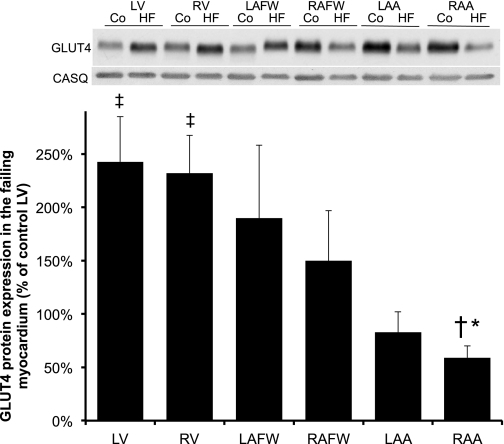
Fig. 2.
Chronic-progressive heart failure (HF) induced alteration in GLUT4 protein expression throughout all cardiac regions. Top: Western blot of cardiac muscles from control (Co) and HF subjects. Top and bottom bands showed a representative immunoblot for GLUT4 (53 kDa) and its corresponding CASQ (60 kDa), respectively. Bottom: mean ± SE of total GLUT4 protein content in dogs with chronic-progressive HF (n = 5). Values represent the content normalized to each respective cardiac chamber. *P ≤ 0.05 vs. failing LV; ‡P ≤ 0.05 vs. control LV; †P ≤ 0.05 vs. failing RV.

