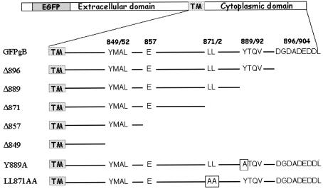
FIG. 1.
Representation of HSV-1 gB cytoplasmic domain. The top drawing depicts the EGFP-tagged gB. The line below depicts the cytoplasmic domain and the locations of highly conserved sorting motifs. The amino acid numbers bracketing the motifs are shown above. The bottom seven lines show the cytoplasmic domains of the mutated gB forms used in the study. Altered amino acids are shown in shaded boxes.

