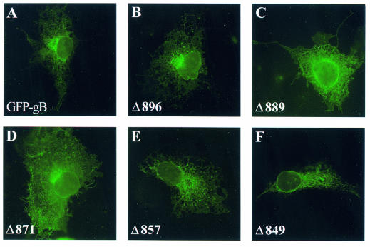
FIG. 2.
Comparative localization of GFP-gB and the truncated forms in transfected cells. Cos-7 cells were transfected with pGFP-gB or each of the truncated forms. Images show spontaneous GFP fluorescence 24 h after transfection. Fluorescence was visualized with a Zeiss Axiophot conventional microscope.

