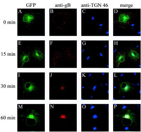
FIG. 5.
Internalization of GFP-gB. Cos-7 cells expressing GFP-gB were incubated at 4°C with an anti-gB antibody for 1 h and then returned to 37°C for 0 min (A to D), 15 min (E to H), 30 min (I to L), or 60 min (M to P). Cells were fixed in methanol and labeled with an anti-TGN 46 antibody. Images show spontaneous GFP fluorescence (A, E, I, and M) and indirect immunofluorescence from the anti-gB antibody and a Cy3-labeled secondary antibody (B, F, J, and N) or from the anti-TGN 46 antibody and Cy5-labeled secondary antibody (C, G, K, and O). (D, H, L, and P) Merged images. Fluorescence was visualized with a Bio-Rad MRC 1000 confocal microscope.

