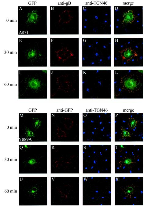
FIG.6.
Internalization of Δ871 and Y889A. The same experiment as that for Fig. 5 was reproduced in Cos-7 cells expressing the truncated Δ871 protein or the mutated Y889A gB protein. Subcellular localization was examined by confocal microscopy as described in the legend for Fig. 5. (A to D and M to P) 0-min incubation at 37°C; (E to H and Q to T) 30-min incubation at 37°C; (I to L and U to X) 60-min incubation at 37°C. EGFP fluorescence of Δ871 and of Y889A is shown in panels A, E, and I and panels M, Q, and U, respectively; anti-gB staining of Δ871 and of Y889A is shown in panels B, F, and J and panels N, R, and V, respectively; and anti-TGN 46 staining of Δ871 and of Y889A is shown in panels C, G, and K and panels O, S, and W, respectively. (D, H, L, P, T, and X) Merged images.

