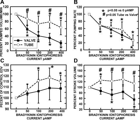
Fig. 6.
Chronotropic and mechanical responses of tubular and bulb regions to bradykinin iontophoretic application. A: pumped volume/min is analogous to cardiac output calculated from frequency and stroke volume. B: the frequency of pumping decreased essentially identically for tubular and valvular regions. C: end-diastolic volume (EDV) increased significantly for the tubular regions and, as shown in D, stroke volume significantly increased for the tubular regions but significantly decreased for the valve regions as bradykinin release was increased. The combination of data as pumped volume/min indicated a substantial drop in valve region flow due to the combination of decreased frequency and stroke volume. A much smaller decrease in flow occurred in the tube region due to increased stroke volume to offset the decline in contraction frequency. *Significant change from control. #Significant difference between valve and tube regions.

