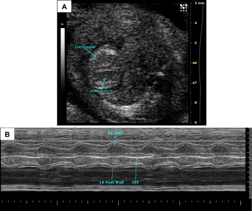
Fig. 10.
Embryonic day (E)15.5 mouse embryonic heart. A: in embryonic stages, blood produces a very echogenic (bright) signal with high-frequency ultrasound (contrast to hypoechoic in the adult). The ventricles can be seen in this image. B: M-mode image of E16 mouse embryo showing myocardial walls, interventricular septum, and ventricles. y-axis shows depth (in mm), and x-axis shows time (in s). Images were acquired using the Vevo 2100 ultrasound system (Visualsonics).

