Fig. 4.
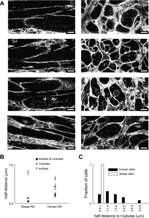
T tubules are present in human atrial cells. A: example of deconvolved 2-μm-thick images of human atrial cell membranes from 4 patients in tissue sections labeled with WGA. T tubules are clearly present in some cells and absent in others in the plane shown. Images at left are longitudinal in orientation while those at right are transverse. None specific staining of the nucleus was observed in some cells an example of which is circled at right. All scale bars are 10 μm and are located at bottom right. B: half distance of voxels within the cell to membrane for sheep and human right atrial cells; surface membrane is grey triangles, t-tubular membrane is dark grey circles, and both membranes together are black squares. C: frequency histogram displaying binned values for the half distance to t tubules for human atrial cells (filled bars) and sheep atrial cells (open bars).

