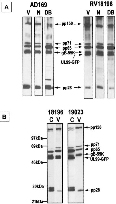
FIG. 5.
Immunoblot analysis of VPs and infected cell proteins with HCMV VP polyclonal antiserum. Extracellular VPs were separated by using a sucrose gradient. (A) AD169 and RV18196 extracellular VPs; (B) RV18196- and RV19023-infected cell and virion proteins. V, virions; N, NIEPs; DB, dense bodies; C, infected cell proteins.

