Fig. 1.
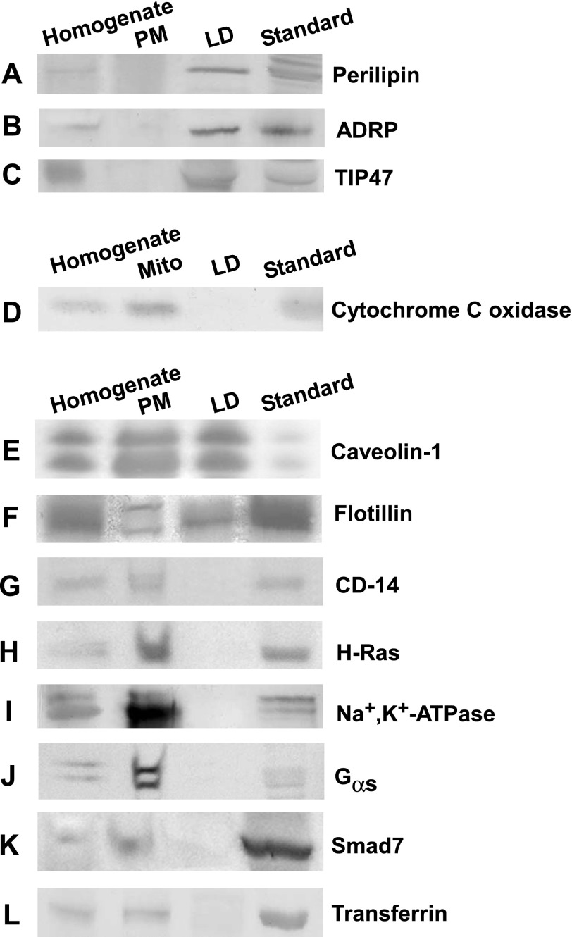
Western blot analysis of adipocyte homogenate, plasma membrane (PM), and lipid droplet (LD) fractions. The relative enrichment of LD, mitochondrial, and PM proteins in isolated adipocyte fractions was determined from Western blots loaded as follows: adipocyte homogenate (lane 1), PM (lane 2), LD (lane 3), and protein standards (lane 4). Proteins present in LD but not PM fractions included perilipin (A), adipose differentiation-related protein (ADRP; B), and tail-interacting protein 47 (TIP47; C). D: proteins in mitochondrial fractions but not LD included cytochrome c oxidase. Proteins in both LD and plasma membrane fractions included caveolin-1 (E) and flotillin (F). Proteins in plasma membrane but not LD fractions included CD-14 (G), H-ras (H), Na+,K+-ATPase (I), Gsα (J), Smad7 (K), and transferrin (L). The protein standard in A–C was detected from homogenates of mouse adipose tissue. Standard in D–L was detected in homogenates of mouse liver.

