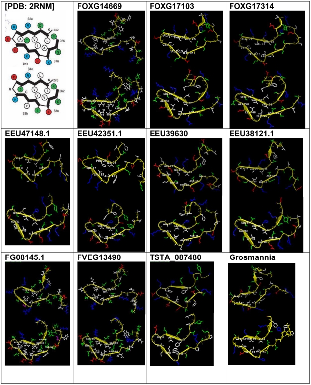
Figure 4. Models of HET-s homologs with structural homology to the HET-s PFD.
The original solenoid [PDB: 2RNM] [6], [7] is shown in the top left corner. Ten structural homologs are represented, including the small s protein homolog from G. calivigera. The structure of FG10600.1 has been shown by Wasmer et al [10] and is not included here. For each structure, two rungs for each solenoid are represented, with the first rung at the top. Amino acids are color-coded as follows: acidic (Asp, Glu) in red, basic (Arg, Lys, His) in blue, nonpolor (Met, Phe, Pro, Trp, Val, Leu, Ile, Ala) in white, polar (Ser, Thr, Asn, Gln, Tyr) in green, and the protein backbone in yellow.

