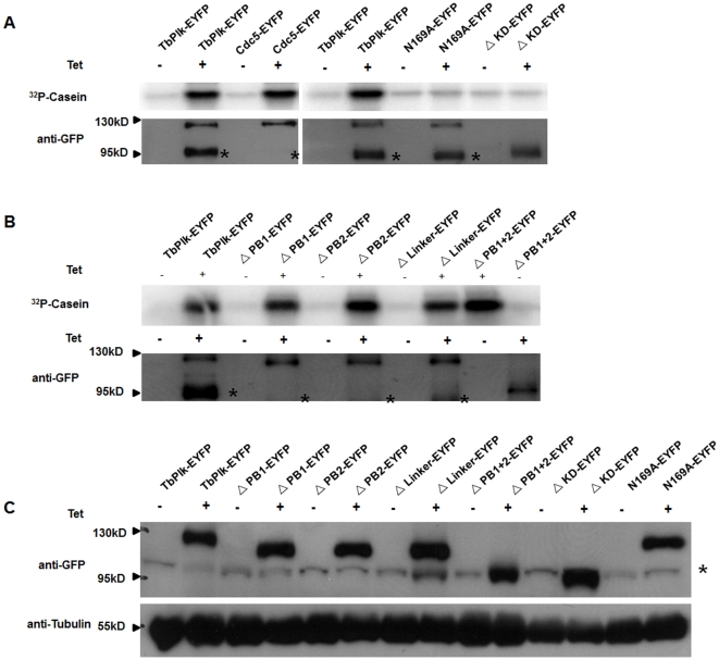
Figure 3. Assay of kinase activities in Cdc5-EYFP, TbPLK-EYFP and the mutants of TbPlk-EYFP.
(A) The kinase activity of Cdc5-EYFP, TbPlk-EYFP, ΔKD-EYFP and N169A-EYFP. (B) The kinase activity of ΔPB1-EYFP, ΔPB2-EYFP, ΔLinker-EYFP, ΔPB1+2-EYFP mutants. All the fusion proteins were immunoprecipitated from the cell lysate with anti-GFP beads. The immunoprecipitates were quantified in Western blots and each incubated with 10 µg dephosphorylated casein and 5 μCi [γ-32P]ATP at 37°C for 1 hr. (C) Crude cell lysates of the transfected cell lines were analyzed on a Western blot stained with anti-GFP antibody. α-tubulin was stained with anti-tubulin antibody and used as a loading control. All the studies were derived from three independent experiments. The asterisks next to the Western blot indicate a nonspecific protein band.

