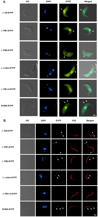
Figure 4. Localization of TbPlk-EYFP mutants in the transfected T. brucei cells and the cytoskeleton isolated from them.
(A) Procyclic-form 29-13 cells expressing individual TbPlk-EYFP mutants were each cultured for 1 day in the presence of 1.0 µg/ml tetracycline, fixed with 3.7% formaldehyde, stained with DAPI and examined with fluorescence microscopy. (B) The cytoskeletons of the transfected cells were isolated after detergent and high salt treatment. They were stained with the L3B2 antibody to FAZ (red) and DAPI for DNA (blue) and observed with fluorescence microscopy. The results were from three independent experiments. Arrows indicate the signals of TbPlk-EYFP mutants. Bars: 2 µm.

