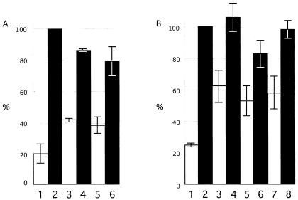
FIG. 4.
Kinase assay to evaluate phosphorylation of IE63 mutants expressed in 293T cells. 293T cells were transfected with the pLXIN-based ORF63 mutants designed to substitute alanine for S or T residues that were putative phosphorylation targets, or with control plasmids, and harvested in RIPA buffer after 36 h. IE63 was immunoprecipitated with polyclonal rabbit antiserum, and IE63 phosphorylation by cellular S/T kinases was assessed with radiolabeled ATP, followed by SDS gel electrophoresis and phosphorimager detection. The bar graph in panel A shows the results of three independent kinase assays performed with the multiple putative phosphorylation site mutants and reported as a mean percentage relative to intact IE63 (bar 2). The other bars are 1, negative control (pLXIN), 3, CompletePhos− mutant, 4, 5′Phos− mutant, 5, CenterPhos− mutant; and 6, 3′Phos− mutant. Panel B shows the combined results of three independent kinase assays performed with the mutants disrupting single putative phosphorylation targets relative to intact IE63 (no. 2); the other bars are 1, negative control (pLXIN), 3, S165, 4, T171, 5, S173, 6, S181, 7, S185; and 8, S186. The lines indicate standard errors.

