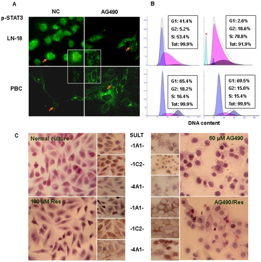
Figure 6. Sensitivities of LN-18 and PBC cells to STAT3 inhibitor AG490.
(A) Immunofluorescence illustration of intracellular distribution of STAT3 in LN-18 and PBC cells without (N) and with (AG490) 60 µM AG490 treatment. Arrows indicate the cells shown in the insets in high magnification (X400). (B) Flow cytometry analysis revealed reduction of G1-phase cells, accumulation of S-phase cells and induction of apoptosis (blue peak) in AG490-treated LN-18 cell population. The cell cycle progression was almost unchanged in PBC cells. *, indicates the peak of apoptotic cells. (C) H&E morphologic examination of LN-18 cells under normal culture or incubated with 100 µM trans-resveratrol, 60 µM AG490 or resveratrol/AG490 mixture for 48 hours (Main images). Small images: immunocytochemical illustration of SULT1A1, 1C2 and 4A1 expression in LN-18 cells treated by 60 µM AG490 without/with 100 µM resveratrol supplementation. Normally cultured and 100 µM resveratrol-treated LN-18 cells were cited as controls.

