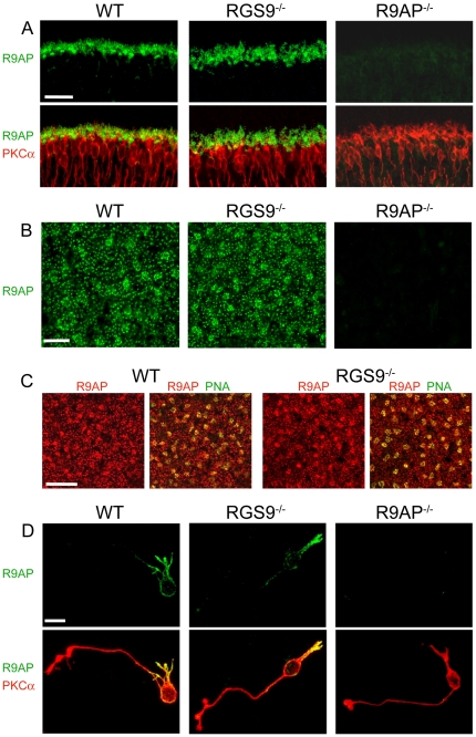
Figure 1. R9AP expression in ON-bipolar cells of WT and RGS9 −/− retinas.
(A) Co-immunostaining of retina cross-sections from WT, RGS9 −/− and R9AP −/− mice for R9AP (green) and the rod ON-bipolar cell marker, PKCα (red). Shown are the outer plexiform layers of each section. Scale bar: 25 µm. (B) Single confocal sections through the outer plexiform layers of whole mount retinas of WT, RGS9 −/− and R9AP −/− mice immunostained for R9AP. We observed no notable differences in the pattern of R9AP immunostaining between RGS9 −/− (40 frames from 6 retinas) and WT controls (30 frames from 6 retinas). Scale bar: 10 µm. (C) Single confocal sections through the outer plexiform layers from whole mount retinas of WT and RGS9 −/− mice co-immunostained for R9AP (red) and peanut agglutinin (PNA, green) labeling cone pedicles in the outer plexiform layer. Scale bar: 25 µm. (D) Rod ON-bipolar cells dissociated from WT, RGS9 −/− and R9AP −/− retinas co-immunostained for R9AP and PKCα. Scale bar: 10 µm.

