
Figure 1. Generation of γ-HIF1α-M3 transgenic lines.
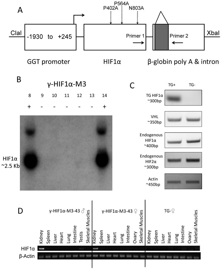
A. Construction of γ-HIF1α-M3 plasmid. Three mutations (P402A, P564A, N803A), dashed arrows. Intron (shaded square) in β-globin poly-A. Primers 1, 2 amplify the transgene by RT-PCR. B. Southern Blot of some TG+ and TG- Founders. Founders #8 and #14, TG+; others are TG-. C. HIF1α transgene, endogenous VHL, HIF1α, and HIF2α RT-PCR γ-HIF1α-M3-43 kidneys. HIF1α transgene, detected only in TG+. Endogenous VHL, HIF1α, and HIF2α mRNAs are expressed at similar levels in TG+ and TG-. β-Actin, loading control. D. HIF1α transgene RT-PCR in multiple organs of γ-HIF1α-M3-43 mice. HIF1α transgene, detected specifically in γ-HIF1α-M3-43 TG+ kidneys. No transgene expression in any organ of TG- mice. β-Actin, loading control.