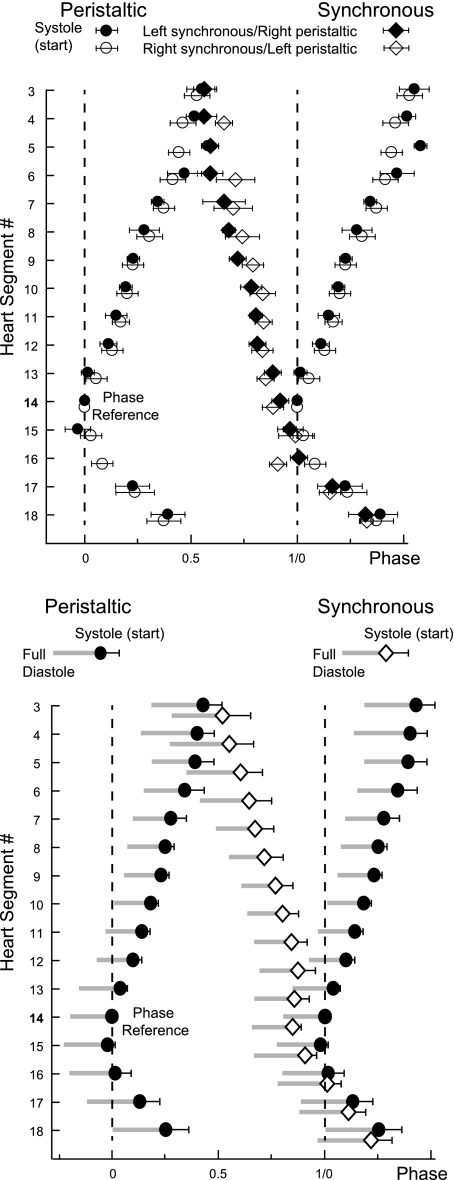
Fig. 2.
Phase diagrams of the constriction patterns of heart segments 3 to 18 from an individual, intact leech (top) and of the average from 13 different preparations (bottom). Vertical dashed lines are provided to facilitate observation of relative phasing. Both phase diagrams show the start of systole (±SD; systole in short) in the peristaltic (circles) and in the synchronous mode (diamonds). Phase reference is heart segment 14 on the peristaltic side, and its systole is assigned 0 phase. To illustrate the intersegmental phase differences in the rear, the peristaltic side is duplicated and shifted by 1. Top: mean phase (±SD) of systole of 9–19 heart beats was determined in an individual juvenile leech. Filled symbols represent 1 switch cycle (left synchronous/right peristaltic); open symbols represent another switch cycle (right synchronous/left peristaltic). Phases could not always be determined in all 32 segments in both switch cycles; for example, segment 16 in the peristaltic mode was obtained once in this animal. Note that patterns are similar on both body sides. Cycle period: 4.1 s. The number of beats per switch cycle was 20 and 21, respectively. Bottom: average of the start of systole and additionally that of full diastole (left edge of gray bars) is shown. Note that heart segments in the front and in the rear converge in phase and that rear heart segments 16 to 18 on both sides fill (diastole) and empty (systole) from front to rear. Graph shows the average (±SD) of the average of the 2 switch cycles analyzed per preparation. Mean cycle period: 3.9 ± 0.45 s.

