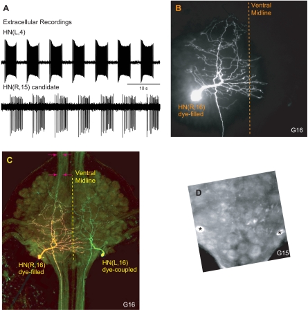
Fig. 3.
Identification and neuroanatomy of additional HN interneurons. A: extracellular recordings from the HN(L,4) interneuron and from a neuron in segment 15 on the contralateral side [HN(R,15) candidate]. Note that burst activity in these 2 neurons is time-locked. B and C: ventral aspect of a dye-injected HN(R,16) interneuron. Anterior is up. B: the confocal image shows rhodamine dextran in the injected cell. The HN(R,16) interneuron has a main neurite that loops and gives rise to many secondary neurites. The main neurite tapers in diameter to form a rearward-going axon leaving the ganglion. The anterior-going neurites do not leave the ganglion. Note that some processes cross the ventral midline. C: superimposed confocal image shows both rhodamine dextran (red fluorescence) and the Alexa fluorophore coupled to Neurobiotin (green fluorescence). The color combination makes the injected cell appear yellow because it contains both rhodamine dextran and Neurobiotin. Its contralateral homolog, the HN(L,16) interneuron, appears green due to dye coupling. Note the 2 neurites in the anterior right connective (arrows). D: same preparation, ganglion 15. Two cell bodies in the position of the HN(15) interneurons (posterior lateral packet) are labeled with Neurobiotin (asterisks).

