Fig. 1.
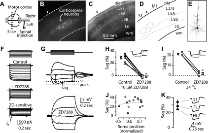
Corticospinal neurons express high levels of hyperpolarization-activated current (Ih). A: injection of fluorescent beads into the cervical spinal cord resulted in retrograde labeling of neurons (B) in the contralateral motor cortex. C: bright-field image and (D) diagram showing cortical layers (L) in motor cortex (wm, white matter; M1, motor cortex; S1, somatosensory cortex). Arrowheads, border between somatosensory cortex (to the left) and M1 (to the right). E: 2-photon image of a biocytin-filled corticospinal neuron in the motor cortex. F: voltage-clamp mode recording (voltage steps: multiples of ±10 mV, holding potential −50 mV) from a corticospinal neuron eliciting currents before (Control) and after (+ZD7288) application of ZD7288 (25 μM; 34°C). Current sensitive to ZD7288 (ZD; bottom) represents Ih. G: current-clamp recording from a corticospinal neuron showing sag of the membrane potential to a steady-state (ss) level and sensitivity to the Ih blocker ZD7288 (25 μM; current steps: multiples of ±50 pA; 22°C). H: ZD7288 sensitivity at 10 μM. Data points for individual neurons are connected by lines, flanked by group means (n = 7; bars: ±SE, *P < 0.05, paired t-test; 22°C). I: sag and sensitivity to ZD7288 measured at 34°C, 25 μM ZD7288 (n = 3; *P < 0.05, paired t-test). J: sag was independent of somatic sublayer location (normalized distance: pia = 0; white matter = 1; line, linear regression; 22°C). K: average (±SE) sag for corticospinal neurons in forelimb (FL; n = 49) and hindlimb (HL; n = 10) regions of the motor cortex and in somatosensory cortex (n = 10).

