Fig. 2.
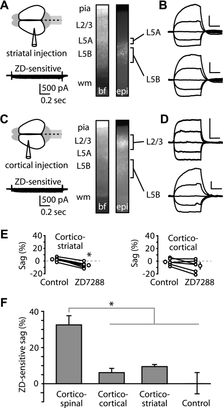
Crossed corticostriatal and corticocortical neurons express low levels of Ih. A: tracer injection into the contralateral dorsolateral striatum resulted in retrograde labeling of crossed corticostriatal neurons in motor cortex, distributed in layers 5A and 5B (images: bf, bright field; epi, epifluorescence). Bottom: ZD7288-sensitive current from a recording in voltage-clamp mode (voltage steps −80 to +10 mV in 10-mV increments; holding potential −50 mV; 34°C) of a layer 5A corticostriatal neuron. B: current-clamp recordings from labeled corticostriatal neurons in layers 5A and 5B. Scale bars: 10 mV, 0.2 s (applies throughout). C: injection into the contralateral motor cortex labeled callosally projecting corticocortical neurons in motor cortex, distributed across multiple superficial and deep layers. Bottom: ZD7288-sensitive current from a voltage-clamp mode recording (voltage steps same as A) of a layer 5B corticocortical neuron. D: recordings from layers 2/3 and 5 corticocortical neurons. E: effect of ZD7288 on sag in corticostriatal (n = 6; *P < 0.05, paired t-test; 22°C) and corticocortical neurons (n = 6; P > 0.05, paired t-test; 22°C). F: ZD7288-induced change in sag for corticospinal (n = 7), corticocortical (n = 6), and corticostriatal (n = 6) neurons (*P < 0.05, t-test, corticospinal vs. other groups). Control neurons (n = 3) are time-control recordings from corticospinal neurons with no drug application to test for Ih changes over the duration of the recording.

