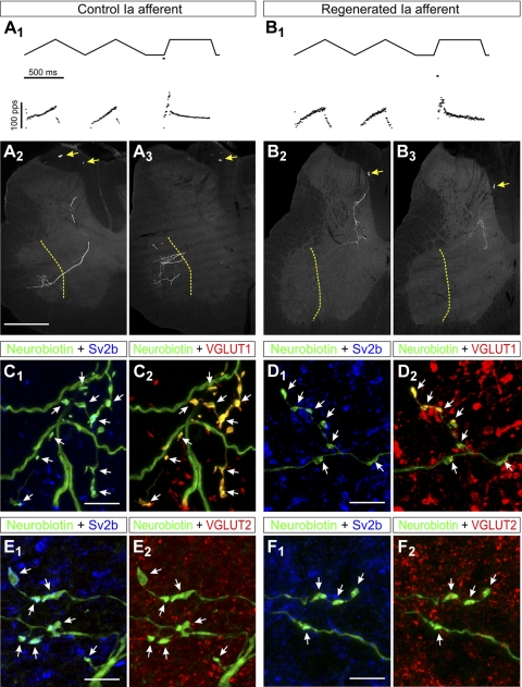
Fig. 10.
Trajectories and VGLUT immunoreactivities of IA afferent fibers in control animals or after peripheral nerve injury and regeneration. A1 and B1: responses of control (A1) and regenerated (B1) sensory afferents to triangular and ramp and hold muscle stretches. The muscle stretch stimulus is shown in the top traces, and the recorded responses depicted as firing frequency time plots are shown in the bottom traces. Both fibers faithfully encode muscle stretch parameters with dynamic responses typical of IA sensory afferents, similar levels of static response during hold phases, and similar history dependence of the initial burst in triangular stretches. Stretch responses were indistinguishable in control and regenerated afferents. A2 and A3: low-magnification epifluorescence images of 2 semiserial spinal cord sections containing labeled collaterals of the sensory axon with stretch responses shown in A1. Segments of the parent axons are visible in the dorsal columns (arrows) and collaterals with terminal branches in laminae V, VII, and IX are easily observed (border between laminae IX and VII is labeled with a dashed yellow line). The arborization in lamina IX was consistently very profuse in all control afferents. B2 and B3: images similar to A2 and A3 but for labeled collaterals from a regenerated axon. Note the parent axon in the dorsal column (arrows) and similar dorsomedial to ventrolateral trajectories of the central collaterals innervating the spinal cord, but these stop before entering lamina IX. C: high-magnification confocal microscopy of a varicose collateral from a neurobiotin-filled IA afferent (FITC-streptavidin, green) with several boutons (arrows) containing immunoreactivity for the synaptic protein SV2b (C1, blue, Cy5) and VGLUT1 (C2, red, Cy3). There was always a perfect correspondence between SV2b-containing varicosities and VGLUT1. D: similar high-magnification images of varicosities (arrows) from a regenerated afferent. All varicosities contained Sv2b (D1) and VGLUT1 (D2); however, neurobiotin-filled varicosities (FTIC, green in D1 and D2) appear of smaller size than in control uninjured afferents. E and F: similar sequence of images but for sections immunolabeled with SV2b and VGLUT2. SV2b-IR neurobiotin-filled varicosities (arrows) in control (E1) and regenerated (F1) afferents lack visible VGLUT2 immunoreactivity (E2 and F2). Scale bars, 500 μm in A1 (A2, B1, and B2 are at the same magnification); 10 μm in C1, D1, E1, and F1 (C2, D2, E2, and F2 are at the same magnification).

