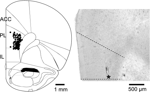
Fig. 1.
Histologically verified recording sites of 46 neurons in the prelimbic cortex (PL). Diagram (adapted from Paxinos and Watson 1998) shows coronal brain section at 3.2 mm anterior to the bregma. Symbols indicate locations of recording electrode tips based on electrolytic lesions. ACC, anterior cingulate cortex; IL, infralimbic cortex. Histology image on the right shows an individual example of a lesion site in the PL (indicated by an asterisk) in 1 brain slice stained with hematoxylin and eosin.

