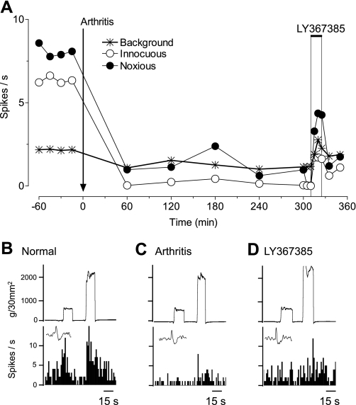
Fig. 6.
mGluR1 antagonist partially reverses inhibition of a mPFC neuron in arthritis pain. Extracellular recordings of the responses of 1 mPFC neuron to brief (15 s) innocuous and noxious stimulation of the knee and background activity. A: time-course data show the decrease in activity after arthritis induction and the partial reversal by a selective mGluR1 antagonist [(S)-(+)-α-amino-4-carboxy-2-methylbenzeneacetic acid (LY367385), 1 mM; concentration in microdialysis fiber, 15 min] administered into the mPFC. ACSF was administered as vehicle control before (predrug) and after LY367385. B–E: peristimulus time histograms show action potentials (spikes)/s. Insets show individual action potentials. Top traces are recordings of the force (g/30 mm2) applied to the knee joint.

