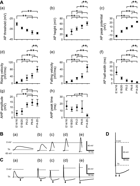
Fig. 5.
Developmental changes in electrophysiological properties of AP and after-hyperpolarization (AHP). A: changes in AP (a–f) and AHP (g and h) properties. Values of AP threshold (Th) and AP peak potential are means ± SE, and other values are medians within the 25th–75th percentile range. Statistical differences in AP threshold and AP peak potential between groups were tested using Scheffe and Games-Howell tests, respectively, and the differences in other values between groups were tested using Mann-Whitney tests. Horizontal bars above each graph indicate pairs with significant differences; *P < 0.01 and **P < 0.05. B: representative traces to illustrate AP shape at different ages. C: representative traces showing AHPs at different ages. The peak of AHP is indicated by an arrowhead below each trace. D: peaks of fast (filled arrowhead) and slow (open arrowhead) AHPs in a neuron at P14–20.

