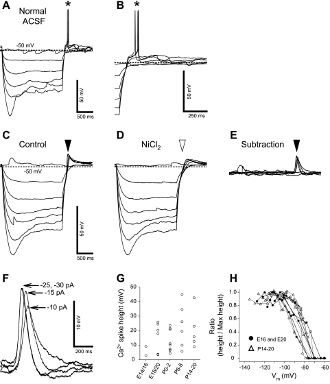
Fig. 7.
Postinhibitory rebound (PIR) and low voltage-activated (LVA) Ca2+ spike in a neuron at E18. A: membrane depolarizations and AP spikes (asterisk) induced by PIRs after membrane hyperpolarizations, which were evoked by negative current injection steps (0–30 pA in 5 pA step, 2-s duration). ACSF, artificial cerebrospinal fluid. B: termination of hyperpolarizations of A with expanded time scale. C: membrane depolarizations induced by PIRs in the presence of TTX (filled arrowhead). D: membrane depolarizations in the presence of NiCl2 and TTX. Transient membrane depolarizations were eliminated by NiCl2 (open arrowhead). E: Ni2+-sensitive spikes (filled arrowhead) computed by subtraction of traces in D from those in C, indicating LVA Ca2+ spikes. F: expanded voltage and time scale of traces in E. Intensities of hyperpolarizing currents are indicated on the right of each trace of the Ca2+ spikes. G: relationship between Ca2+-spike amplitudes and different age groups. H: relationships of hyperpolarized Vm with Ca2+-spike amplitude in prenatal (circles) and postnatal (triangles) neurons. The spike amplitudes were normalized to the maximum amplitude.

