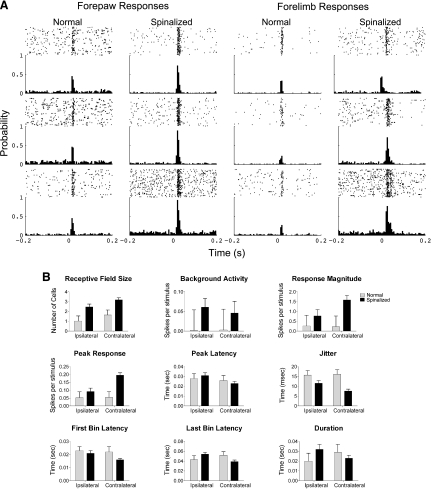
Fig. 2.
A: peristimulus time rasters and histograms generated from cells simultaneously recorded from the hindlimb sensorimotor cortex during stimulation of the cutaneous forelimbs under light anesthesia. The responses of 6 representative cells are shown from each group of rats. Each cell responded to passive stimulations of the cutaneous forepaw digit 2. The PSTH of cells recorded from the spinalized rats had greater spontaneous activity, greater response magnitudes relative to the spontaneous activity, and their peak latency was shifted in time but the duration of their response was not significantly different from the normal rats. The rasters above each PSTH show the spikes (dots) per trial (row of dots). B: response measures of cells in the hindlimb somatosensory cortex recorded during passive sensory stimulations of the cutaneous forelimbs separated by whether the stimulus was ipsilateral or contralateral to the neuron for spinalized rats and normal rats: receptive field size, background activity, response magnitude, peak response, latency of the peak of the response, jitter to the first response, first bin latency, last bin latency, and duration of the response.

