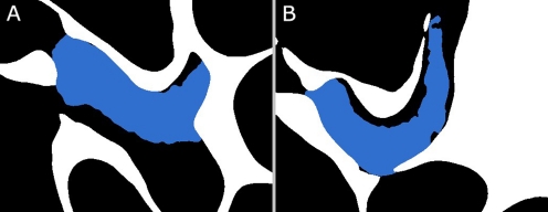
FIG. 12.
Cross sections at different depths through the 3D merged models of the bulla bone (white) from μCT and the tensor tympani (blue) from OPFOS. Black represents air-filled space such as the ME air cavity. The tensor tympani fits nicely in the bone, rather touching the cavity wall than overlapping with it.

