
Fig. 1.
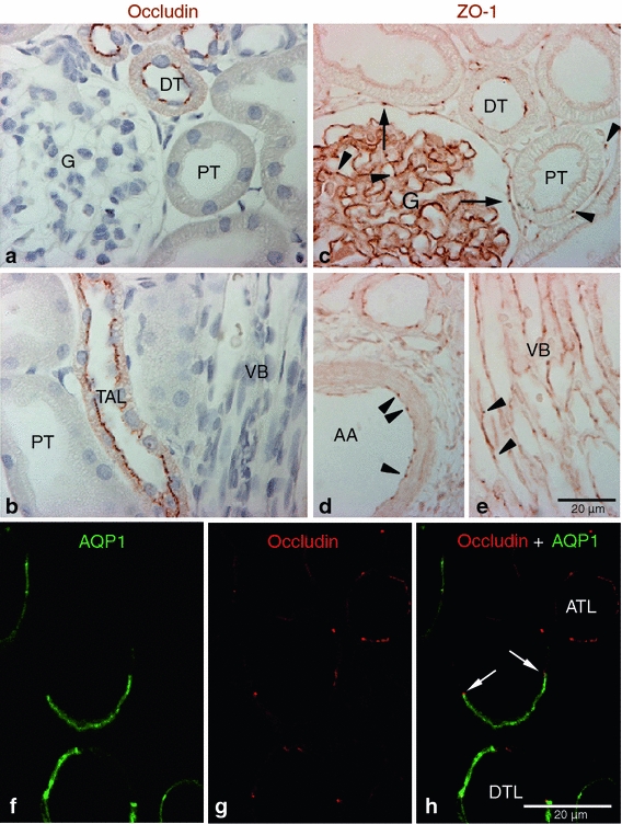
Occludin and ZO-1 expression in control kidney. Representative of occludin (a, b), ZO-1(c–e), and occludin-AQP1 confocal (f–h) immunostaining images obtained from sham-operated control kidneys. Occludin (brown) immunoreactivity was observed as dot-and-line patterns at the apical end of the lateral membrane in tubular epithelial cells in the cortex (a) and outer medulla (b). Occludin was expressed primarily in the distal tubule (DT) including the thick ascending limb (TAL). No occludin immunolabel was detected in glomeruli (G), proximal tubule (PT), or blood vessels, including the vascular bundles (VB). ZO-1 (brown) immunoreactivity was observed in all tubular segments, including the proximal tubule (PT) and distal tubule (DT) (c–e). ZO-1 labeling also appeared in the glomerular parietal epithelial cells (arrows) and in endothelial cells (arrowheads) of all blood vessels including the glomerular capillary, peritubular capillary, arcuate artery (AA), and vascular bundle (VB). To identify the thin limbs of the loop of Henle, confocal immunofluorescence staining for occludin (red) and AQP1 (green) were performed (f–h). Occasionally, transition points (white arrows) from the AQP1-positive descending thin limb (DTL) to AQP1-negative ascending thin limb (ATL) were observed and occludin immunoreactivity appeared in both thin DTL and ATL segments (g, h). Results are representative of findings in the five control rat kidneys