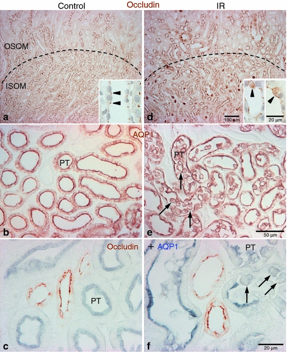
Fig. 2.

Effects of IR injury on occludin immunolabel in the rat kidney. Representative micrographs showing occludin (a, d), AQP1 (b, e) and occludin-AQP1 double immunostaining (c, f) images in the outer medulla in sham-operated control (a–c, n = 5) and IR (d–f, n = 5) kidneys. Dashed lines indicate boundary between the outer stripe of the outer medulla (OSOM) and inner stripe of the outer medulla (ISOM). Compared with control kidneys, abnormal localization of occludin (brown) immunoreactivity was observed in the outer medullary region in ischemic kidneys (d). In a subpopulation of cells, occludin diffusely localized in the cytoplasm (inset, arrowheads) in the ischemic kidneys (d). Many AQP1 (brown in b, e; blue in c, f)-positive proximal tubular (PT) cells lost polarity, detached from basement membrane, and were present in the tubule lumen (arrows) in ischemic kidneys (e, f). Occludin (brown in c, f) immunoreactivity was not observed in the proximal tubule either under control or following IR injury
