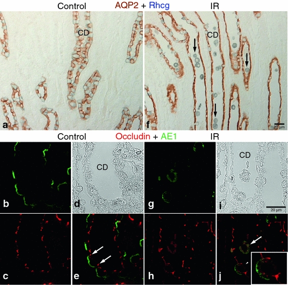
Fig. 4.

Effects of IR injury in the OMCD. Representative of AQP2-Rhcg double immunohistochemistry (a, f) and occludin-AE1 confocal fluorescent microscopy (b–e, g–j) images in the outer medulla in sham-operated control (a–e, n = 5) and IR (f–j, n = 5) kidneys. Compared with control, many Rhcg (blue)-positive collecting duct (CD) cells detached from the basement membrane and were present in the tubule lumen (arrows) in ischemic kidneys (f). However, AQP2 (brown)-positive cells were not damaged after IR injury. Confocal images confirmed the intercalated cell-specific damage in the CD. In control kidneys, occludin (red) labeling was observed at the apical end of the lateral membrane in both principal and intercalated cells (e, white arrows). In IR injury kidneys, both occludin and AE1 (green) lost their normal polarity and were localized diffusely in the cytoplasm in cells released into the lumen (j, white arrow). Partial delocalization or internalization of occludin and AE1 was also observed in cells still remaining in the tubule wall (white arrowheads, inset) in ischemic kidneys (j)
