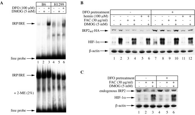
FIG. 8.
Conditional inhibition of iron-mediated decrease in IRP2 expression by DMOG. (A) DMOG has no apparent iron-chelating properties. B6 and parent H1299 cells were either left untreated (lanes 1 and 4) or treated for 6 h with 5 mM DMOG (lanes 2 and 5) or 100 μM DFO (lanes 3 and 6). Cytoplasmic extracts from B6 (10 μg) or H1299 (30 μg) cells were analyzed for IRE-binding activity with a 32P-labeled IRE probe in the absence (top) or presence (bottom) of 2% 2-mercaptoethanol (2-ME). The positions of the IRE-IRP complexes and excess free IRE probe are indicated by arrows. (B) Effects of DMOG on steady-state levels of chimeric wild-type IRP2. H1299 cells were incubated in the absence of tetracycline for 2 days to express chimeric wild-type IRP2. Half of the cells were left untreated, and the other half received 100 μM DFO overnight to further stimulate IRP2 expression. Tetracycline was then added back to the medium to shut off IRP2 expression, and the cells were either left untreated or pretreated with 5 mM DMOG for 2 h. Subsequently, the cells were either left untreated (lanes 1, 2, 7, and 8) or treated in the presence or absence of 5 mM DMOG for 4 h with 30 μg of FAC/ml (lanes 3, 4, 9, and 10) or 100 μM hemin (lanes 5, 6, 11, and 12). Lysates were analyzed by Western blotting with an antibody against HA (top), HIF-1α (center), or β-actin (bottom). (C) Effects of DMOG on steady-state levels of endogenous IRP2. Parent H1299 cells were either left untreated (lanes 1 to 3) or pretreated overnight with 100 μM DFO (lanes 4 to 6). The cells were then either left untreated (lanes 1 and 4) or treated for 4 h with 30 μg of FAC/ml in the presence (lanes 3 and 6) or absence (lanes 2 and 5) of 5 mM DMOG. The inhibitor was administered 2 h prior to addition of FAC. Lysates were analyzed by Western blotting with an antibody against IRP2 (top), HIF-1α (center), or β-actin (bottom).

