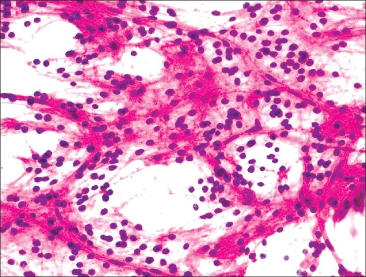
Figure 5.

Dysembryoplastic neuroepithelial tumor: Microcysts separated by clusters of neurocytic cells with rounded bland nuclei, in a sparse meshwork of fine fibrillary neuropil-like processes. Large ganglion cells are noted embedded in the wall of microcysts (H and E, ×400)
