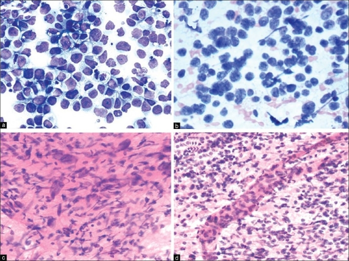
Figure 7.

(a) Germinoma: Syncytial arrangement of loosely cohesive neoplastic cells with vacuolated cytoplasm, moderate nuclear pleomorphism, prominent nucleoli and frequent mitoses. Occasional interspersed mature lymphocytes are noted (H and E, × 200). (b) Medulloblastoma: Sheets of malignant cells with primitive appearing nuclei, attempted Homer-Wright rosettes and fine neuropil-like intercellular material (H and E, ×400). (c) GBM: Markedly pleomorphic malignant astrocytic cells in well-formed fibrillary meshwork (H and E, ×200). (d) GBM: Malignant small astrocytic cells in well-formed fibrillary meshwork with a hyperplastic blood vessel (H and E, ×100)
