FIG. 2.
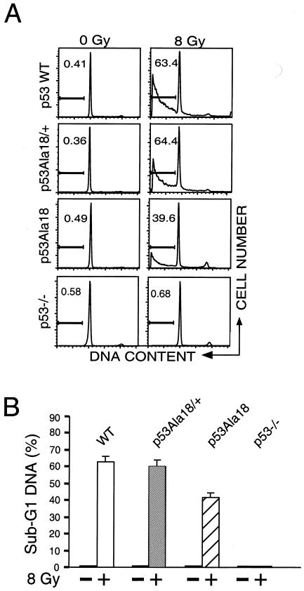
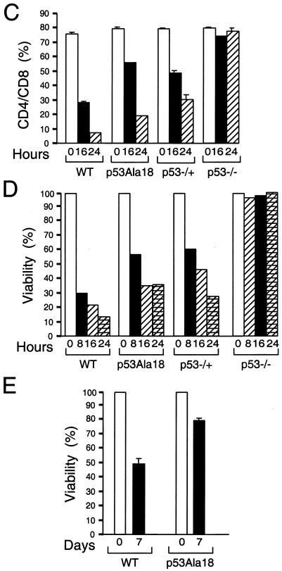
p53-mediated apoptosis is defective in p53Ala18 mice. (A) Analysis of sub-G1 content of thymocytes removed from irradiated and unirradiated animals (8 Gy, 8 h posttreatment). The y axis depicts cell number, and the x axis represents DNA content. The percentage of sub-G1 cells is given for each sample. WT, wild type. (B) Histograph of sub-G1 DNA from irradiated and nonirradiated mice. Two independent animals were analyzed in triplicate for each genotype. (C) Time course of CD4+ CD8+ profile of mice irradiated (8 Gy) in vivo and nonirradiated mice. The graph depicts double-positive cells. (D) Viability of thymocytes over time in response to ionizing radiation. Thymocytes were removed from mice, irradiated (8 Gy), and harvested at various times (0, 8, 16, and 24 h). Cells were stained with annexin V-fluorescein isothiocyanate, anti-CD4+, and 7AAD. Values given are the averages of the viable population (annexin V-fluorescein isothiocyanate and 7AAD negative) and are normalized to the number of viable cells in the untreated animal for each genotype. (E) Apoptosis in splenocytes is reduced in p53Ala18 mice. Splenocytes were prepared and plated (2 × 10⁵ cells). Cells were irradiated with 8 Gy. Cell viability was determined 7 days post-ionizing radiation. The x axis represents time (days), and the y axis represents viability relative to unirradiated control cells.


